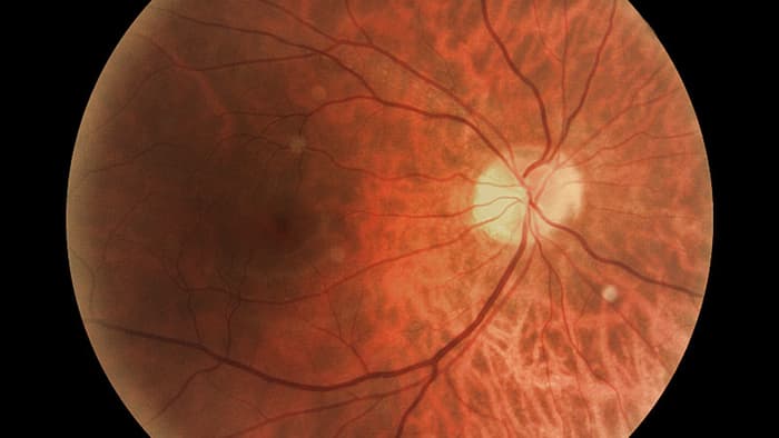
Eyes are complex machines, and visual processing of the images they take in accounts for more than a third of brain functioning. It starts with photoreceptor cells at the back of the retina that react to light wavelengths and send their electrically-coded data to the 30 or so types of retinal ganglion cells, each one specializing in processing specific aspects of vision; they're also the only nerve cells connecting the eye with the brain.
Projected from the ganglion cells are long, slender projections called axons, which are bundled together along the optic nerve from the eye before fanning out to various regions in the brain where the visual input is interpreted. Unfortunately, axons in the brain or spinal cord don't regenerate on their own once they've been damaged, leading to permanent vision loss. One reason for this is the reduction over time of a cascade of growth-enhancing molecular interactions within axon cells known as the mTOR pathway.
The condition of the mice's eyes in the study was similar to glaucoma, which is associated with pressure on the optic nerve to the point where damage occurs. Affecting nearly 70 million people globally, glaucoma and is the second-leading cause of blindness after cataracts, and currently there's no cure. Injuries, retinal detachment, and some tumors and brain cancers can also damage the optic nerve.
For the study, adult mice with the optic nerve crushed in one eye were put through one of two regimens: daily exposure to a moving black and white grid that offered high-contrast stimulation, or biochemical manipulations that stimulated the mTOR pathway within the ganglion cells. Each approach produced a modest regrowth of axons, though not long enough to reach and communicate with their specific destinations in the brain.
But when both therapies were used together, while a blinder was placed on the mouse's good eye, a significant number of axons grew enough to reach their specific target in the brain to process that visual input, which allowed the mouse to partially see with its previously blind eye. To test it, the researchers showed the mouse an expanding dark circle that resembled an approaching predatory bird, which caused it to run for cover.
But the mice failed other visual tests that probably required finer visual discrimination, according to one of the study's authors, Andrew Huberman, associate professor of neurobiology at Stanford University. The next step in the team's research is to try and expand the number of axons that reach their destination in the brain, as well as assess the 30 or so subtypes of retinal ganglion cells.

 Previous page
 Back to top


