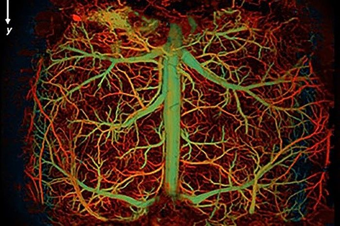
Scientists
at Duke University have developed an ultrafast photoacoustic imaging system
capable of capturing the functional and molecular changes that occur in major
brain disorders.
Imaging techniques that provide real-time, detailed information in relation to complex cerebral vascular networks are important for expanding our understanding of neurovascular disorders like stroke, dementia, and acute brain injury.
While positron emission tomography (PET) and functional magnetic resonance imaging (fMRI) provide decent images, they suffer from low spatial resolution, making it difficult to differentiate between adjacent bodily structures, and low temporal resolution, which is the time it takes to yield measurements and construct an image.
Similarly, optical microscopy produces high-resolution images but is hindered by slow imaging speed and poor penetration depth. Microbubble-enhanced ultrasound penetrates deeply with high resolution but lacks functional sensitivity.
An alternative method of imaging, photoacoustic microscopy (PAM), uses pulses of laser light fired into an organ. The pulses cause an ultrasound wave which is captured to form an image.
Importantly, PAM can use laser light of different wavelengths to target specific structures in the body, even down to the molecular level. This means that PAM can measure important hemodynamic parameters such as blood oxygenation, blood flow, and metabolic rate of oxygen.
The downside to PAM is that it is slow to scan. But this problem has been solved by Duke Institute of Brain Sciences (DIBS) researchers with the development of ultrafast functional photoacoustic microscopy (UFF-PAM) that is two times faster than existing PAM systems.
UFF-PAM enables the imaging of brain microvasculature and functioning with a wide field of vision and high spatial resolution that is lacking in other imaging techniques.
In a proof-of-concept experiment, Duke researchers used UFF-PAM to successfully capture hemodynamic responses to induced hypoxia, sodium nitroprusside-induced hypotension, and stroke in mouse brains. UFF-PAM was able to capture rapid, whole-brain changes in real time.
The stroke experiment yielded an unexpected result. UFF-PAM detected a spreading depolarization (SD) wave emanating from the area of the stroke across the brain, causing narrowing of blood vessels (vasoconstriction) as it spread. SD waves are of great interest to researchers and scientists because their function is poorly understood.
“SD waves could be an indication of the level of severity of an injury, making them a potential diagnostic tool,” said Junjie Yao, PhD, assistant professor of biomedical engineering and DIBS faculty member.
“The nature of the waves could also offer clues to the type and extent of brain injury, which could inform and optimize treatment,” Yao said.
The team at Duke is now looking at using UFF-PAM to study other diseases. While UFF-PAM is currently only being used in animals, Yao revealed plans to develop a handheld UFF-PAM for use on humans.
The study appeared in the journal Light: Science and Applications.

 Previous page
 Back to top


