Complex cognition and behavior,
in animals and humans alike, hinges on information flowing across a network of
deeply interconnected brain cells. For scientists, the scale of that network
posed a major obstacle to better understanding the mechanics of cognition,
because available imaging tools were historically incapable of tracing how
neurons fire in sync from the far reaches of the cortex. That need gave rise to
the idea of a "mesoscope"—an imaging technology with microscopic
resolution fine enough to resolve single cells, but a macroscopic field of view
large enough to capture neurons across broad swathes of the brain.
Now, a new study in Nature Methods describes a mesoscopic technique that may allow scientists to plumb the breadth and depth of the brain at peak resolution, scale, and speed. "The challenge with using mesoscopes for visualizing the fast activity of single neurons in 3D is that high-resolution point-scanning approaches are typically needed, for which the scanning time scales very unfavorably with the size of the imaged volume," says Rockefeller University's Alipasha Vaziri.
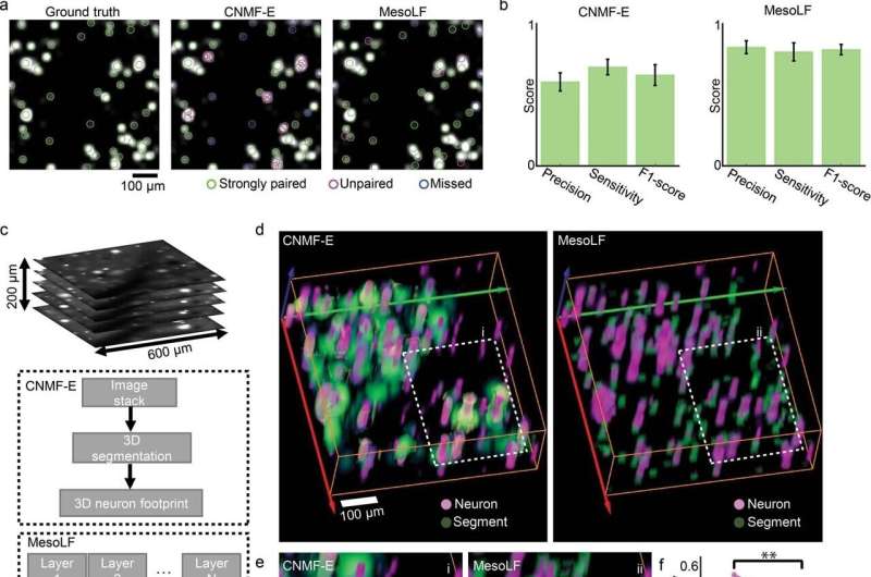
The technology, dubbed MesoLF, can capture key interactions between 10,500 neurons distributed with a volume in the mouse brain at once—imaging cells buried at previously inaccessible depths, firing from brain regions many millimeters apart, simultaneously—with unprecedented resolution.
MesoLF is a spin-off of light-field microscopy (LFM), a 3D imaging technique known for providing fast, high-resolution imaging. For all its strengths, however, LFM performs poorly deep inside scattering tissue such as the mouse brain, where dense tissue scatters light.
Vaziri previously circumvented some of these limitations with a machine-learning algorithm developed by his team, which estimates the locations of active neurons to better detect brain cell activity in dense tissue. His latest work expands that reach by adding software and hardware to scale up the system, allowing it to peer into tissues of various shapes and rigidities. Crucially, it also keeps the computational costs inherent to processing terabytes of raw data as low as possible.
"This is made possible through a custom optical design for maintaining high optical imaging resolution over mesoscopic volumes, in combination with a set of algorithmic innovations that scale our modular computational pipeline's capacity and capabilities accordingly," Vaziri says.
Given the relatively low cost barrier in optical hardware, Vaziri hopes to make his MesoLF technique widely available to scientists studying the inner workings of the brain. His designs are now available under an open-source license.

 Previous page
 Back to top


