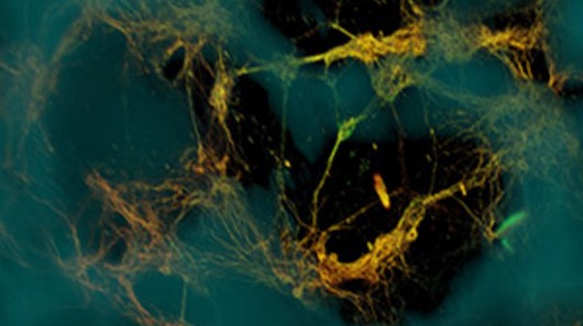
One approach to studying the brain rather than working on the whole thing at once is to examine small bits of it. With that in mind, researchers at the Tissue Engineering Resource Center at Tufts University, Boston have developed a three-dimensional brain-like tissue that is structurally similar to living rat brain tissue, functions enough like it for experimental purposes, and is able to be kept alive for up to two months.
One approach to studying the brain rather than working on the whole thing at once is to examine small bits of it. With that in mind, researchers at the Tissue Engineering Resource Center at Tufts University, Boston have developed a three-dimensional brain-like tissue that is structurally similar to living rat brain tissue, functions enough like it for experimental purposes, and is able to be kept alive for up to two months.
The advantages of tissue testing is that it's much easier and precise than working with entire organs, its more humane than experimenting on animals, and the experiments can be tailored to suit the problem. The stumbling block is making the tissue to suit the job, and growing neurons in Petri dishes doesn't always meet the need.
Cells grown in culture don’t cooperate in re-creating the structures found in living organisms because they only grow in two dimensions, don’t segregate properly into white (axons) and grey matter (neuron cell bodies), don’t generate the complex structures found in living brains, and tend not to live very long. It is possible to grow neural tissues in three dimensions, but the cells don’t naturally produce the right structures in the laboratory. They need to be guided, and that is where the Tufts team comes in.
The Tufts' solution was to work out a way to three dimensionally grow tissue that shows complex structure, properly segregates white and grey matter, and responds like proper brain tissue. The team did this by using two materials; a silk protein, which is used to build a scaffolding; and a collagen-based gel.
The scaffolding holds the material together and the gels encourage the cells to grow through it – much like ants in the jelly of the more high-tech varieties of ant farms you find in toy stores. It’s made by building a donut out of the silk protein in which the scientists mix rat neurons. In the middle of the donut is, appropriately, the "jelly" made of the collagen protein, which oozes into the scaffolding.
As the neurons grow, they form networks around the pores of the scaffolding and axon projections grow into the center to connect to the opposite side. The end result is a structure that is very similar to proper neural tissue that lives a lot longer than the 2D Petri dish variety and shows a similar gene expression to that of growing nerve cells.
The new tissue lives up to two months and the scientists found that it shows electrical activity and responsiveness similar to that in a living brain, and that it responds to injuries and drugs in a similar fashion.
The team believes that this new tissue would be very useful for studying brain trauma and the effects of drugs on the neural system. In fact, they've already had a go at dropping weights on the tissue and found that it responded in ways similar to that of living animals. The scientists say that, aside from ethical concerns, the tissue is superior to animals for study because the tissue can be studied immediately after trauma instead of waiting for dissection and preparation, and it’s possible to track healing in the tissue over extended periods of time.
In the future, the team says that this artificial tissue could be used to help develop new treatments for brain dysfunction, but, for now, the next step for the team will be to replace the donut scaffolding with six concentric circles with each containing a different type of neuron to mimic the six layers in the human cortex.
"Good models enable solid hypotheses that can be thoroughly tested," says Rosemarie Hunziker, Ph.D., program director of Tissue Engineering at the NIH’s National Institute of Biomedical Imaging and Bioengineering (NIBIB). “The hope is that use of this model could lead to an acceleration of therapies for brain dysfunction as well as offer a better way to study normal brain physiology.” The team’s findings were published in Proceedings of the National Academy of Sciences.

 Previous page
 Back to top


