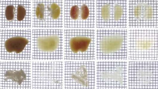
Japanese researchers have found a way to turn tissue transparent in mice, allowing them to see cellular networks and gain a better understanding of biological systems. Researchers say this may ultimately lead to deeper comprehension of autoimmune and psychiatric diseases given it can assist in 3D modelling of organs including the brain.
How do you improve a mouse? Researches at RIKEN Quantitative Biology Center and the University of Tokyo have tried a new approach: make it see-through. This breakthrough enables very detailed shots of the creature’s individual organs or the entirety of the organism to be taken. This is, say researchers, “the ultimate dream of systems biology” and will bring them closer to understanding how life works, by being able to watch almost from the inside out.
The technique is based on a brain imaging method called CUBIC (Clear Unobstructed Brain Imaging Cocktails and Computational Analysis). The method centers around chromophores, a molecular subunit in the body that absorbs light. One kind of chromophore, heme, which forms part of haemoglobin, is found in most of the body's tissues. The researchers discovered that the amino alcohols in the CUBIC reagent stopped the heme from blocking light as it generally does. Once stopped organs became “dramatically more transparent.”
Building on this technique the scientists were able to get 3D models of organs whilst they remain in the body by taking slices using light-sheet microscopy. They took slices of varied organs of mice and found clear differences in the pancreas of diabetic and non-diabetic mice.
Although this technique still cannot be used on living things, the process is still considered very useful for understanding the 3D structure of organs given its extremely precise, single-cell imaging. “It allowed us to see cellular networks inside tissues, which is one of the fundamental challenges in biology and medicine," says one the paper's authors , Kazuki Tainaka.
So what are the useful applications? Plenty, according to the researchers. “It could be used to study how embryos develop or how cancer and autoimmune diseases develop at the cellular level,” say lead researcher Hiroki Ueda. Ultimately this may lead to a better understanding of the human brain, given the precise imaging, and it may ultimately lend greater understanding to psychiatric or autoimmune diseases. However there is still much work to be done, say the researchers.
The research was recently published in the journal Cell

 Previous page
 Back to top


