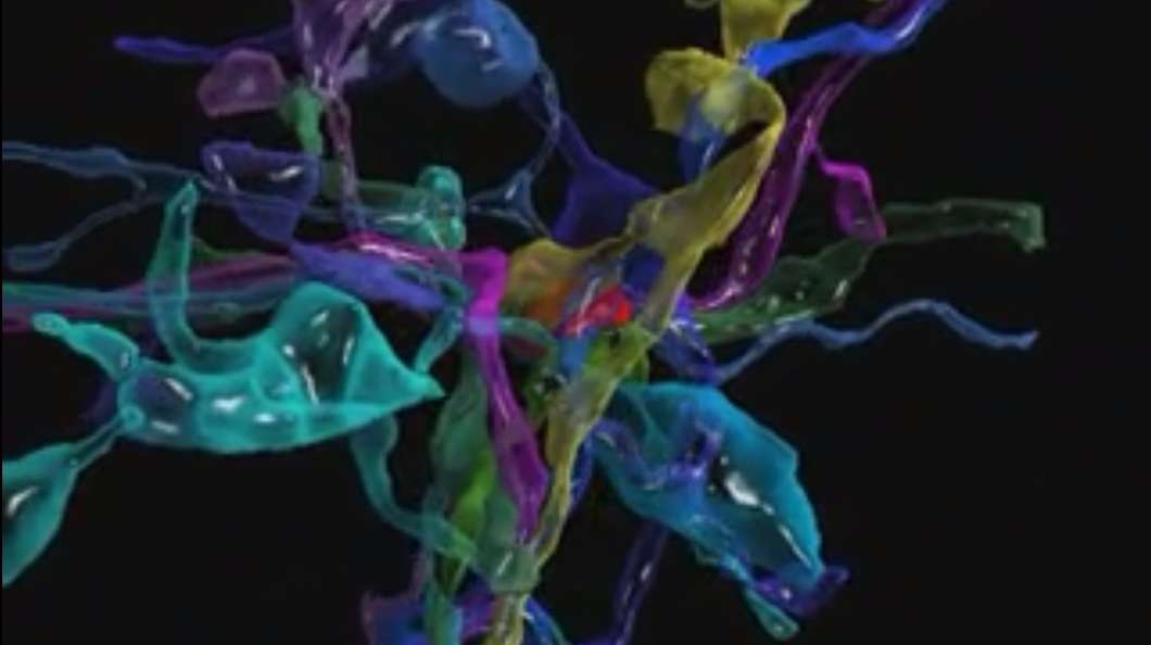
The human brain contains more synapses than there are galaxies in the observable universe (to put a number on it, there are perhaps 100 trillion synapses versus 100 billion galaxies), and now scientists can see them all – individually. A new imaging tool promises to open the door to all sorts of new insights about the brain and how it works. The tool can generate images at a nanoscale resolution, which is small enough to see all cellular objects and many of their sub-cellular components (so for the biology-literate, that's stuff like neurons and the synapses that permit them to fire, plus axons, dendrites, glia, mitochondria, blood vessel cells, and so on).
Developed by researchers at the Boston University School of Medicine and Harvard University, the imaging method employs an automated tape-collecting device equipped with a diamond knife to obtain ultra-thin brain sections, which are then scanned under an electron microscope. Different colors are used to identify different cellular objects using software developed by study co-author Daniel Berger.
To demonstrate their new tool the researchers peered inside the brain of an adult mouse. They imaged a very small piece of a mouse's neocortex at a resolution that made individual synaptic vesicles visible (these are tiny spheres of less than 40 nm diameter that store neurotransmitters, or chemical signals, for release from a synapse into a "target" neuron). The specific area they imaged is involved in receiving sensory information from mouse whiskers, which are much more sensitive than human fingertips.
Noting significant redundancies in the synaptic connections, the researchers analyzed how axons and dendrites connect. An axon is a nerve fiber; it transmits electrical impulses like a fiber optic cable. You can think of dendrites as being like the input sockets on your electronics; they're the branches that jut out from a neuron, receive the impulses from axons, and convert that information into a signal the neuron understands.
It had previously been thought that the connectivity between axons and dendrites could be inferred from their locations – a concept called Peters' Rule, despite the fact that the man responsible for the idea disputes it. The researchers proved this is not the case. There is a more complex relationship between axons and dendrites, which causes multiple synapses to form on some dendrites but not on others. The best predictor of synapse formation turns out to be the presence of another synapse between that same axon and dendrite pair.
The researchers take this as evidence that imaging at super-fine detail will be necessary for neuroscientists to fully understand the brain. On the downside, it seems that more data is going to mean more questions and not necessarily more insights, but the researchers draw an optimistic conclusion from this – "there is no reason to stop doing it until the results are boring," they write.
Further, study first author Narayanan "Bobby" Kasthuri says that the complexity of the brain is far greater than anyone had imagined. "We had this clean idea of how there's a really nice order to how neurons connect with each other," he explains, "but if you actually look at the material it's not like that. The connections are so messy that it's hard to imagine a plan to it. But we checked, and there's clearly a pattern that cannot be explained by randomness."
Nanoscale imaging of the brain means that in every square micrometer of tissue we can see things we've never seen before. It promises huge opportunities for discovery and exploration, and it promises that someday, in the far flung future, we may have our answers – we may learn everything there is to learn about how the brain works.
Many scientists see the work as a waste of time and money, senior author Jeff Lichtman notes – it is simply too enormous an undertaking to yield much value. But surely we as a species must continue to dream big, to chase the impossible, because history has taught us that the journey, the striving for more answers, proves as rewarding and fruitful as the goal that we may never reach.

 Previous page
 Back to top


